Are you experiencing back pain or discomfort? Perhaps you’ve heard the term “disc protrusion” mentioned by your healthcare provider, but you’re not quite sure what it means or how it is diagnosed. In this article, we will guide you through the diagnosis process for disc protrusion, providing valuable insights into the steps involved and the various medical professionals you may encounter along the way. By understanding this process, you can take an active role in your healthcare and ensure that you receive the best possible treatment for your condition.

What is Disc Protrusion?
Disc protrusion is a medical condition that affects the shock-absorbing discs between the vertebrae of your spine. When a disc protrudes, it means that the outer layer of the disc has bulged or weakened, causing it to press against nearby nerves or other spinal structures. This can lead to pain, numbness, and other symptoms. Disc protrusion is also commonly referred to as a herniated disc or a slipped disc.
Symptoms of Disc Protrusion
The symptoms of disc protrusion can vary depending on the location and severity of the condition. Common symptoms include back or neck pain, tingling or numbness in the arms or legs, muscle weakness, and difficulty with coordination or balance. You may also experience shooting pain that radiates down your arm or leg, known as radicular pain. In severe cases, disc protrusion can lead to bladder or bowel dysfunction, which requires immediate medical attention.
When to Seek Medical Attention
If you are experiencing persistent or worsening back or neck pain, along with any of the symptoms mentioned above, it is important to seek medical attention. Additionally, if you have sustained a recent injury or have a history of spinal conditions, it is advisable to consult a healthcare professional. Ignoring the symptoms of disc protrusion can lead to further complications and may delay appropriate treatment.

The Role of Medical History
When you visit a healthcare professional for disc protrusion, they will take a detailed medical history to understand your symptoms, previous injuries, and any underlying conditions. Your medical history can provide valuable information about the possible causes and contributing factors of your disc protrusion. It can also help your healthcare provider develop a suitable treatment plan and determine the most appropriate diagnostic tests to confirm the diagnosis.
Physical Examination
During a physical examination, your healthcare provider will assess your range of motion, muscle strength, reflexes, and any specific areas of tenderness or pain. They may also perform specialized tests, such as the straight leg raise test for assessing nerve irritation. This examination helps your healthcare provider localize the source of your symptoms and rule out other potential causes of your pain.
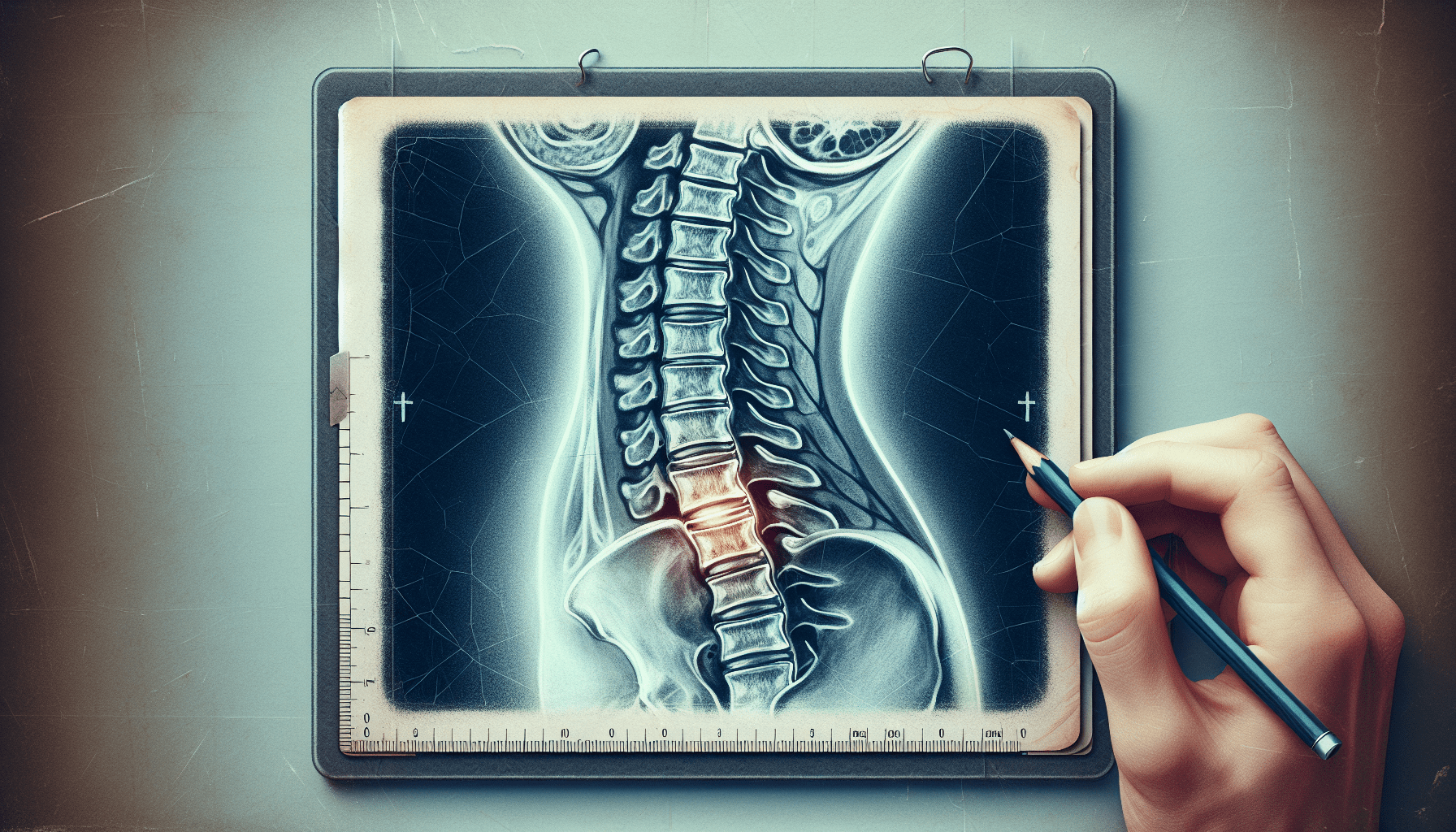
Diagnostic Imaging
To confirm the diagnosis of disc protrusion, various diagnostic imaging tests may be ordered. Imaging tests provide a detailed view of the spine, allowing healthcare professionals to identify the specific location and severity of the disc protrusion. The most commonly used imaging tests for disc protrusion include X-rays, MRI scans, and CT scans.
Types of Diagnostic Imaging
X-rays
X-rays are a common initial imaging test to evaluate the spine. They can provide a clear image of the bones, showcasing any abnormalities or changes in the structure of the spine. While X-rays cannot directly visualize the discs or nerves, they can help rule out other conditions and provide important information about the overall health of your spine.
MRI Scans
Magnetic Resonance Imaging (MRI) scans are more detailed and can provide a comprehensive view of the discs, nerves, and surrounding tissue. This imaging test uses powerful magnets and radio waves to create detailed images of the spine. MRI scans are highly effective in diagnosing disc protrusion and can help determine the appropriate treatment approach.
CT Scans
Computed Tomography (CT) scans are often used in conjunction with MRI scans to gain a more complete understanding of the extent and location of the disc protrusion. CT scans use X-ray technology combined with computer processing to create cross-sectional images of the spine. This allows healthcare professionals to assess the structure and alignment of the vertebrae and identify any potential complications or additional factors contributing to your symptoms.
Interpreting Diagnostic Imaging Results
Interpreting the results of diagnostic imaging can be complex and requires the expertise of a healthcare professional. Radiologists are specialized doctors who interpret imaging tests and provide detailed reports to your healthcare provider. They will analyze the images produced by the X-rays, MRI scans, or CT scans and provide a diagnosis based on their findings. Your healthcare provider will then review the results with you, explaining the implications and discussing potential treatment options.
Additional Tests
In some cases, additional tests may be necessary to further evaluate the extent of your disc protrusion or to assess other potential causes of your symptoms. These tests may include electromyography (EMG) to evaluate nerve function, discography to assess the integrity of the discs, or nerve conduction studies to measure the speed of nerve impulses. Your healthcare provider will determine if any additional tests are needed based on your specific situation.
Treatment Options
The treatment options for disc protrusion can vary depending on the severity of your symptoms and the impact on your daily life. Initially, conservative treatment measures such as rest, physical therapy, and pain medications may be recommended to relieve symptoms and promote healing. In cases where conservative measures are not effective, more invasive treatment options such as epidural steroid injections or surgical intervention may be considered.
Surgery is typically considered a last resort and is reserved for cases where conservative treatments have failed or when severe neurological symptoms are present. The specific surgical procedure will depend on the location and severity of the disc protrusion but may involve removing the protruding disc material or fusing the affected vertebrae together.
In conclusion, disc protrusion is a common spinal condition that can cause significant pain and discomfort. Seeking medical attention and undergoing a comprehensive diagnosis process is crucial in identifying the specific source of your symptoms and developing an appropriate treatment plan. With advancements in diagnostic imaging and a range of treatment options available, healthcare professionals can effectively manage disc protrusion and help you find relief from your symptoms.